💡 Direct Answer & Executive Summary (mRNA to Amino Acid Length Converter)
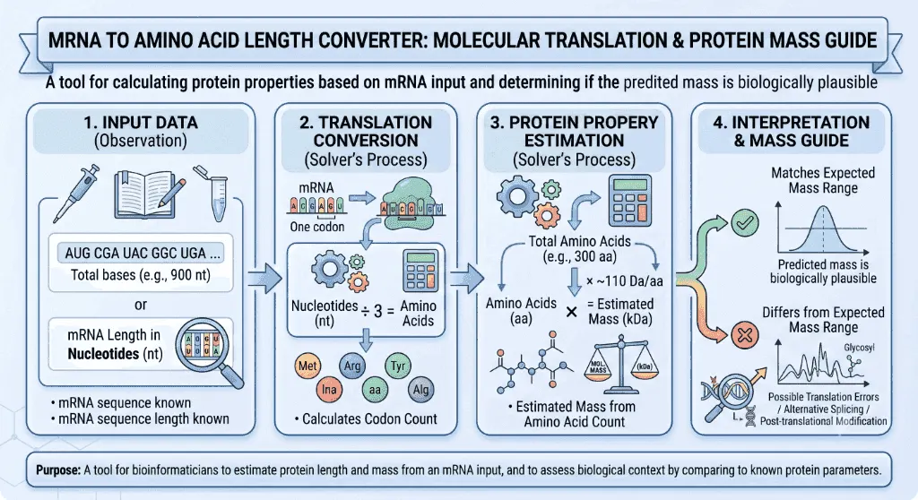
Definition: Convert mRNA nucleotide coding sequence (CDS) length into translated amino acid polypeptide chain length, estimated protein molecular weight (kDa), and ribosomal synthesis time.
Governing Math Formula: Triplet Codon Rule: 3 mRNA nucleotides = 1 amino acid. Protein length: L(AA) = (L_CDS / 3) - 1 (accounting for 1 terminating stop codon). Molecular Weight: MW ≈ L(AA) × 110 Da.
Target Applications: Provides real-time quantitative solutions in Biology for students, engineers, researchers, and finance professionals.
mRNA to Amino Acid Length Converter: Molecular Translation & Protein Mass Guide
1. Introduction
When the first synthetic mRNA vaccines against COVID-19 were engineered in early 2020, molecular bioengineers did not synthesize physical viral proteins by hand. Instead, they designed an exact string of . When injected into human muscle cells, our cellular ribosomes read this digital genetic program, synthesizing a viral Spike glycoprotein composed of exactly with a predicted molecular mass of approximately .
How does a linear sequence of four ribonucleotide bases () translate into three-dimensional molecular machines like hemoglobin, insulin, antibodies, and collagen? Why does an insertion or deletion of just a single nucleotide cause catastrophic diseases like Duchenne Muscular Dystrophy? How do bioinformaticians accurately predict protein size, synthesis duration, and molecular weight from raw cDNA sequences?
The answer lies in the Genetic Code Translation Rules—the fundamental molecular conversion mechanism that bridges genomic nucleic acids and functional proteomic structures.
flowchart LR
    DNA["🧬 Genomic DNA
Coding Gene (Exons)"] -->|"Transcription
(RNA Pol II)"| MRNA["📜 Pre-mRNA / Mature mRNA
5' Cap + 5' UTR + CDS + 3' UTR + Poly-A"]
    MRNA -->|"Triplet Codon Engine
3 Nucleotides = 1 Codon"| RIBO["🧫 Ribosome 80S / 70S
Aminoacyl-tRNA Delivery at A-P-E Sites"]
    RIBO -->|"Peptidyl Transferase"| PROT["🧪 Nascent Polypeptide Chain
L(AA) = (L_CDS / 3) - 1
Mass ≈ L(AA) × 110 Da"]
Whether you are designing expression plasmids in a synthetic biology laboratory, verifying CRISPR gene-editing reading frames, or preparing for advanced exams in biochemistry, understanding the mathematical and structural relationship between mRNA and protein length is indispensable.
2. Definitions
2.1 Simple Everyday Definition
An mRNA to Amino Acid Converter calculates the exact number of amino acids in a synthesized protein based on the length of its coding RNA sequence. Because the cellular machinery reads genetic messages in three-letter "words" called codons, every three RNA bases correspond to one amino acid block in the growing protein chain.
2.2 Formal Technical Definition
In molecular biology, translation conversion is the quantitative mapping of an Open Reading Frame (ORF) or Coding Sequence (CDS) of length (measured in ribonucleotides) to its corresponding mature polypeptide residue count :
Where:
- The division by reflects the triplet, non-overlapping, commaless nature of the universal genetic code.
- The subtraction of accounts for the terminating stop codon (), which binds protein release factors rather than an aminoacyl-tRNA, terminating peptide elongation without adding an amino acid residue.
2.3 Vivid Real-World Analogies
The 3-Letter Morse Code Telegraph:
Imagine a telegraph cable transmitting a continuous string of letters without spaces: AUGAAAUUUCCCGGGUAACCC. The telegraph operator knows the codebook uses strictly 3-letter words. The operator reads AUG (Word 1: Start/Met), AAA (Word 2: Lys), UUU (Word 3: Phe), CCC (Word 4: Pro), GGG (Word 5: Gly), and UAA (Stop / End of Message). The stop command tells the operator to cut the paper ribbon. Even though 18 letters were transmitted, exactly 5 physical word blocks were assembled.
The Modular Lego Blueprint:
Think of an mRNA strand as a strip of building instructions where every 3 color-coded notches specify one snap-on Lego brick. The very last instruction in the manual is a red "Finish" stamp—it closes the manual but is not a physical brick itself.
3. History & Scientific Milestones
Deciphering how nucleic acid sequences direct protein synthesis is universally recognized as one of the greatest scientific triumphs of the 20th century.
timeline
    title Milestones in the Genetic Code & Translation Biology
    1953 : Watson & Crick : Discover DNA double helix structure
    1954 : George Gamow : Proposes 'Diamond Code' triplet hypothesis (4³ = 64 codons)
    1961 : Nirenberg & Matthaei : Poly-U experiment proves UUU codes for Phenylalanine
    1966 : Khorana, Nirenberg & Holley : Completely decipher all 64 triplet codons
    2000 : Ramakrishnan, Steitz & Yonath : Solve atomic 3D structure of the ribosome (Nobel 2009)
2020 : Karikó & Weissman : Modified nucleoside mRNA enables rapid global mRNA vaccines- George Gamow (1954): The theoretical physicist reasoned that because proteins contain , a 1-base code () or a 2-base code () would be mathematically insufficient. A 3-base triplet code () provided more than enough coding capacity to specify all 20 amino acids with built-in redundancy.
- Marshall Nirenberg & J. Heinrich Matthaei (1961): In a historic cell-free experiment at the NIH, they added a synthetic poly-uracil RNA strand (
UUUUUU...) to a bacterial extract and synthesized a polypeptide composed entirely of polyphenylalanine, cracking the very first genetic codon word.
- Har Gobind Khorana & Robert Holley (1966): Synthesized defined repeating RNA copolymers to decode all remaining codons, discovering the start codon () and the three stop signals (), sharing the 1968 Nobel Prize in Physiology or Medicine.
- Venkatraman Ramakrishnan, Thomas Steitz, & Ada Yonath (2000–2009): Solved high-resolution X-ray crystallographic structures of the , , and ribosomal subunits, demonstrating that the ribosome is a ribozyme—its peptidyl transferase active site is catalyzed purely by ribosomal RNA (rRNA), not proteins.
4. Core Concepts & Theoretical Principles
graph TD
    subgraph Transcript_Architecture["Mature mRNA Structure"]
        CAP["5' Methylguanosine Cap"] --> UTR5["5' Untranslated Region (5' UTR)
Kozak Consensus: (gcc)gccRccAUGG"]
        UTR5 --> START["Start Codon (AUG - Met)"]
        START --> ORF["Open Reading Frame (CDS)
Triplets: Codon 1, 2, 3 ... N"]
        ORF --> STOP["Stop Codon (UAA / UAG / UGA)"]
        STOP --> UTR3["3' Untranslated Region (3' UTR)"]
        UTR3 --> POLYA["Poly-A Tail (150-250 Adenines)"]
    end

    subgraph Protein_Output["Translated Polypeptide"]
        N_TERM["N-Terminus (NH₂-Met)"] --> CHAIN["Amino Acid Residues (1 to n)"]
        CHAIN --> C_TERM["C-Terminus (-COOH)"]
    end

    ORF -->|"Ribosomal Decoding"| CHAIN
4.1 Triplet Codons & Degeneracy
The genetic code consists of :
- specify the 20 standard proteinogenic amino acids.
- (, , ) trigger peptide release.
- Degeneracy / Redundancy: Multiple codons can code for the same amino acid (e.g., Leucine and Arginine are each encoded by 6 distinct codons). However, the code is unambiguous—no single codon ever codes for more than one amino acid.
4.2 Reading Frames & The Open Reading Frame (ORF)
Any linear RNA sequence has three potential forward reading frames depending on whether translation begins at base , base , or base :
- Frame 1:
[AUG] [GAG] [AAA] [CUU] ...
- Frame 2:
A [UGG] [AGA] [AAC] [UU...]
- Frame 3:
AU [GGA] [GAA] [ACU] [U...]
The Open Reading Frame (ORF) is the specific continuous nucleotide stretch that begins with an start codon, proceeds in multiples of three, and terminates at an in-frame stop codon.
4.3 Untranslated Regions (UTRs) vs. Coding Sequences (CDS)
Not all nucleotides in an mRNA molecule are translated into amino acids:
- (Leader Sequence): Regulates ribosomal recruitment and translation initiation efficiency via Kozak sequences (eukaryotes) or Shine-Dalgarno motifs (prokaryotes).
- (Coding Sequence): The precise protein-encoding segment between start and stop codons.
- (Trailer Sequence): Contains miRNA binding sites and polyadenylation signals () controlling mRNA stability and localization.
- : A protective tail of that prevents exonucleolytic degradation.
5. Mathematical Formulas & Derivations
5.1 The Length Conversion Formula
To calculate the total number of amino acids in a mature polypeptide () from a given coding sequence length ( in nucleotides):
5.2 Reverse cDNA Length Formula
To find the required cDNA coding sequence length from a known protein size:
5.3 Protein Molecular Weight () Estimation
The molecular mass of a translated protein is estimated by multiplying the number of amino acid residues by the average amino acid molecular weight:
Why per residue?
- The unweighted average molecular weight of the 20 free standard amino acids is .
- When weighted according to natural abundance in globular proteins (smaller residues like Glycine, Alanine, and Leucine occur far more frequently than bulky Tryptophan or Tyrosine), the weighted average free amino acid mass is .
- During ribosomal peptide bond synthesis, every condensation reaction releases one water molecule (, molecular weight ):
5.4 Variable Reference Table
| Parameter | Symbol | Standard Units | Biological Role | Practical Calculation Note |
|---|---|---|---|---|
| Coding Sequence Length | Nucleotides / Base pairs (bp) | Nucleotide count from AUG through Stop | Must be a multiple of 3 | |
| Total Codons | Codons () | Total 3-base reading units () | Includes | |
| Amino Acid Count | Residues () | Total peptide chain length | Excludes non-coding stop codon | |
| Average Residue Mass | Daltons () | Weighted average residue mass () | Used for protein SDS-PAGE mass estimation | |
| Estimated Protein Mass | or | Total unglycosylated polypeptide mass | ||
| Elongation Velocity | Ribosomal translation speed | Eukaryotes: ; Bacteria: |
6. Step-by-Step Computational Walkthrough
Let us calculate the protein properties for the human Tumor Protein p53 () coding sequence:
flowchart TD
    STEP1["Step 1: Input mRNA CDS Length
L_CDS = 1,182 nucleotides"] --> STEP2["Step 2: Calculate Total Triplet Codons
Total Codons = 1,182 / 3 = 394 codons"]
    STEP2 --> STEP3["Step 3: Subtract Terminating Stop Codon
L_AA = 394 - 1 = 393 Amino Acids"]
    STEP3 --> STEP4["Step 4: Estimate Molecular Weight
MW = 393 × 110 Da = 43,230 Da = 43.23 kDa"]
    STEP4 --> STEP5["Step 5: Estimate Translation Synthesis Time
t_euk = 393 / 6 AA/s ≈ 65.5 seconds (1.09 min)"]- Given:- coding sequence length .
- Calculate Total Codons:
- Calculate Polypeptide Length:
- Estimate Predicted Molecular Mass:(Note: On SDS-PAGE gel electrophoresis, p53 runs anomalously high at due to heavy proline phosphorylation, giving it its historic name!)
- Estimate Ribosomal Synthesis Duration:
7. Visual Explanations & The Ribosomal Elongation Cycle
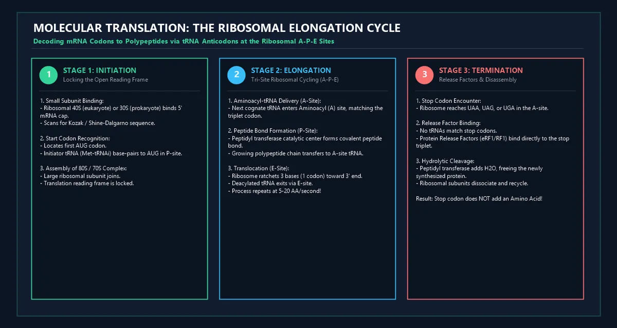
flowchart LR
    subgraph A_Site["1. Aminoacyl (A) Site"]
        A1["Cognate tRNA Enters
Anticodon binds mRNA Codon"]
    end
    subgraph P_Site["2. Peptidyl (P) Site"]
        P1["Peptidyl Transferase Center
Forms Covalent Peptide Bond"]
    end
    subgraph E_Site["3. Exit (E) Site"]
        E1["Deacylated Empty tRNA
Dissociates from Ribosome"]
    end

    A1 -->|"Peptide Transfer"| P1
    P1 -->|"Ribosomal Translocation (EF-G / eEF2)"| E1
graph TD
INPUT{"Input Genomic / Transcript Sequence"}
INPUT --> CHECK_START{"Does Sequence Begin with ATG/AUG?"}
CHECK_START -->|"No"| SCAN["Scan 5' -> 3' for Consensus Kozak / Start AUG"]
CHECK_START -->|"Yes"| CHECK_LEN{"Is Length Divisible by 3?"}
SCAN --> CHECK_LEN
CHECK_LEN -->|"No (Remainder 1 or 2)"| WARN["⚠️ Frameshift / Incomplete Reading Frame"]
CHECK_LEN -->|"Yes (Multiple of 3)"| SCAN_STOP["Scan for In-Frame Stop Codons (UAA, UAG, UGA)"]
SCAN_STOP --> CALC["Apply Formula: L(AA) = (L_CDS / 3) - 1"]
CALC --> OUTPUT["Compute Mass (kDa) and Peptide Sequence"]8. Comparative Analysis & Molecular Reference Tables
8.1 Prokaryotic vs. Eukaryotic Translation Architecture
| Feature | Prokaryotes (Bacteria / E. coli) | Eukaryotes (Human / Yeast / Mammals) |
|---|---|---|
| Ribosome Architecture | ( subunits) | ( subunits) |
| Initiator Amino Acid | -formylmethionine () | Unmodified Methionine () |
| Initiation Signal | Shine-Dalgarno sequence () | scanning + Kozak consensus |
| Transcript Structure | Often Polycistronic (multiple ORFs on 1 mRNA) | Exclusively Monocistronic (1 ORF per mRNA) |
| Translation Velocity | Very rapid: | Moderate: |
| Coupling | Coupled: Transcription and translation occur simultaneously | Separated: Transcription in nucleus; translation in cytoplasm |
8.2 Standard 20 Amino Acid Residue Molecular Mass Reference
| Amino Acid Name | 3-Letter Code | 1-Letter Code | Free Molecular Mass () | In-Chain Residue Mass () | Characteristic Property |
|---|---|---|---|---|---|
| Glycine | $75.07$ | $57.05$ | Smallest, achiral, high conformational flexibility | ||
| Alanine | $89.09$ | $71.08$ | Small, hydrophobic, strong -helix former | ||
| Serine | $105.09$ | $87.08$ | Polar, nucleophilic, primary phosphorylation target | ||
| Proline | $115.13$ | $97.12$ | Rigid cyclic imino acid; creates peptide kinks | ||
| Valine | $117.15$ | $99.13$ | Branched-chain aliphatic hydrophobic | ||
| Threonine | $119.12$ | $101.11$ | Polar hydroxyl group; secondary phosphorylation target | ||
| Cysteine | $121.16$ | $103.14$ | Thiol group; forms covalent disulfide bonds () | ||
| Leucine | $131.17$ | $113.16$ | Highly abundant hydrophobic core residue | ||
| Isoleucine | $131.17$ | $113.16$ | Branched-chain hydrophobic isomer of Leucine | ||
| Asparagine | $132.12$ | $114.10$ | Amide side chain; target for -linked glycosylation | ||
| Aspartate | $133.10$ | $115.09$ | Negatively charged acidic carboxylate () | ||
| Glutamine | $146.15$ | $128.13$ | Polar amide; frequent hydrogen bond donor/acceptor | ||
| Lysine | $146.19$ | $128.17$ | Positively charged basic amine; ubiquitination target | ||
| Glutamate | $147.13$ | $129.11$ | Negatively charged acidic carboxylate () | ||
| Methionine | $149.21$ | $131.20$ | Sulfur-containing; universal Start Codon (AUG) | ||
| Histidine | $155.16$ | $137.14$ | Imidazole ring; physiological buffer () | ||
| Phenylalanine | $165.19$ | $147.18$ | Aromatic benzyl ring; hydrophobic stacking | ||
| Arginine | $174.20$ | $156.19$ | Strongly basic guanidinium group () | ||
| Tyrosine | $181.19$ | $163.18$ | Aromatic phenol; tyrosine kinase phosphorylation | ||
| Tryptophan | $204.23$ | $186.21$ | Largest amino acid; indole ring, absorbs UV at |
8.3 Benchmark Human & Model Organism Proteins
| Protein Name | Biological Function | Coding Nucleotides () | Polypeptide Length () | Observed Molecular Weight |
|---|---|---|---|---|
| Human Insulin (Preproinsulin) | Glucose homeostasis hormone | (Cleaved to ) | ||
| Green Fluorescent Protein (GFP) | Bioluminescent reporter marker | |||
| -Globin (Hemoglobin) | Oxygen transport in erythrocytes | |||
| Human Serum Albumin (HSA) | Colloid oncotic blood pressure | |||
| SARS-CoV-2 Spike Glycoprotein | Viral receptor binding & fusion | (Monomer) | ||
| Human Dystrophin | Muscle sarcolemma structural anchor | |||
| Human Titin | Muscle sarcomere passive elasticity | () |
9. Practical Real-World Applications & Examples
Example 1: mRNA Vaccine Antigen Engineering
In vaccine manufacturing, scientists synthesized the full-length Spike glycoprotein gene of SARS-CoV-2.
- Transcript Coding Length: .
- Codon Count: .
- Translated Residues: .
- Predicted Monomeric Mass: .
Example 2: Recombinant Protein Expression Vector Cloning
A biotechnology team clones the Green Fluorescent Protein (GFP) gene into an E. coli pET-28a expression plasmid.
- GFP ORF Length: .
- Amino Acids: .
- Protein Mass: (Exact amino acid sequence mass: ).
Example 3: Designing a Poly-Histidine Purification Tag (6xHis-Tag)
To purify a target protein via Nickel-NTA affinity chromatography, a molecular biologist adds a tag directly before the stop codon.
- Nucleotides Added: (
5'-CAUCAUCAUCAUCAUCAU-3').
- Additional Mass Added: ().
Example 4: Diagnosing a Frameshift Indel Mutation
A clinical geneticist identifies a single base pair deletion (delG) in exon 3 of a DNA repair gene:
- Because the deletion is (not divisible by 3), all downstream codons are misread in the wrong reading frame.
- After 14 incorrect amino acids, the ribosome encounters a premature stop codon (), yielding a truncated non-functional peptide instead of the wild-type protein.
Example 5: Estimating Proteomic Synthesis Burden in Bioreactors
A industrial fermenter expresses recombinant human albumin ().
- At a translation rate of in engineered bacterial hosts, each individual ribosome takes:
- Polyribosomes (polysomes) with 20 ribosomes per mRNA molecule release one complete albumin molecule every per transcript!
10. In-Depth Case Studies
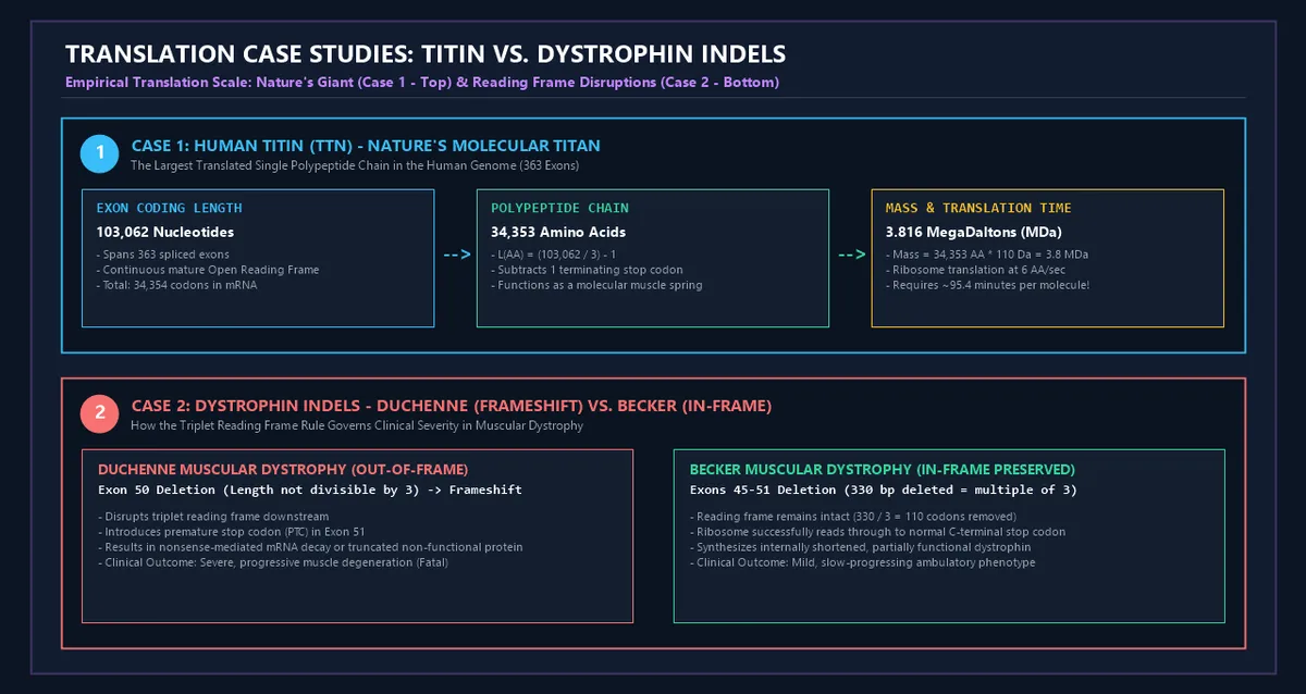
Case Study 1: Human Titin (TTN) — Nature's Giant Polypeptide
- The Biology: Titin is the largest single polypeptide encoded in the human genome, acting as a molecular spring in cardiac and skeletal sarcomeres.
- The Sequence: Spanning 363 exons, its primary canonical isoform transcript has a coding sequence of .
- The Translation: Translates to with a colossal molecular weight of ().
- Translation Duration: In human cardiomyocytes (), a single ribosome requires over 1.5 hours () of continuous translation just to synthesize one single Titin polypeptide!
Case Study 2: Dystrophin Exon Skipping & The Reading Frame Rule
- The Clinical Challenge: Duchenne Muscular Dystrophy (DMD) is caused by deletions in the Dystrophin gene ().
- The Reading Frame Rule: - If an exon deletion disrupts the triplet reading frame (number of deleted bases ), a frameshift introduces a premature stop codon within exons, producing a completely absent or non-functional truncated dystrophin, causing severe, lethal Duchenne DMD.- If a deletion removes a multiple of 3 nucleotides (e.g., in-frame deletion of exons 45–51, total), the reading frame is preserved. The ribosome produces an internally shortened, partially functional dystrophin, resulting in mild Becker Muscular Dystrophy (BMD).
- Therapeutic Breakthrough: FDA-approved antisense oligonucleotide drugs (e.g., Eteplirsen) induce targeted exon-skipping during pre-mRNA splicing to restore the triplet reading frame.
11. Advantages of Sequence-to-Protein Conversion Modeling
- Precise Experimental Planning: Enables molecular biologists to accurately predict band migration on SDS-PAGE, Western blots, and mass spectrometry matrices.
- Rapid Recombinant Construct Design: Instantly verifies that engineered protein tags (, , , ) are correctly in-frame with the primary gene.
- Automated Bioinformatic Pipelines: Powers genome annotation algorithms (such as BLAST, NCBI ORFfinder, and Prodigal) to discover novel protein-coding genes in raw metagenomic sequencing data.
- Biomanufacturing Optimization: Facilitates accurate stoichiometry calculations when formulating mRNA-lipid nanoparticle (LNP) drug doses.
12. Biochemical Complexities & Post-Translational Realities
While and provide reliable baseline estimates, real proteins undergo dynamic modifications:
- Signal Peptide Cleavage: Secreted and transmembrane proteins contain hydrophobic N-terminal signal peptides () that are enzymatically cleaved off by Signal Peptidase in the Endoplasmic Reticulum (ER).
- Post-Translational Modifications (PTMs):- - and -Linked Glycosylation: Addition of complex branched oligosaccharide chains can increase observed molecular weight by (e.g., Erythropoietin glycoprotein).- Phosphorylation ( per phosphate): Modulates protein activity and increases net negative charge.
- Pro-Peptide Proteolytic Processing: Many hormones are synthesized as inactive precursors (e.g., Preproinsulin Proinsulin Mature Insulin chains with C-peptide removed).
- Selenocysteine () and Pyrrolysine (): In specialized selenoproteins, the stop codon is recoded as the 21st amino acid Selenocysteine in the presence of a downstream SECIS RNA hairpin element.
13. Common Mistakes to Avoid
1. Forgetting to Subtract the Terminating Stop Codon:
A common error is dividing by 3 and assuming all codons produce an amino acid. The terminal stop codon () signals peptide termination and does not insert an amino acid residue. Always use .
2. Including $5'$ and $3'$ UTRs or Poly-A Tails in CDS Calculations:
Full mature mRNA transcripts contain several hundred untranslated nucleotides (, , and ). You must isolate only the Open Reading Frame (CDS) between the start and codon before dividing by 3.
3. Assuming Free Amino Acid Mass () Instead of Residue Mass ():
Free amino acids contain an extra water molecule (, ) that is lost during ribosomal condensation. Using overestimates total protein mass by approximately 16%.
12. Frequently Asked Questions (FAQ)
What is the formula to convert mRNA sequence length to amino acid length?
The formula is: Where is the length of the coding sequence in nucleotides.
Why do three nucleotides code for one amino acid?
With four unique RNA bases (), a doublet code can only produce combinations, which cannot cover all 20 standard amino acids. A triplet code provides combinations, sufficient to encode all 20 amino acids with punctuation marks (start and stop signals).
Why is the average molecular weight of an amino acid residue taken as ?
The weighted average molecular mass of free amino acids in natural proteins is approximately . During peptide bond formation, a condensation reaction removes a water molecule (), resulting in an average in-chain residue mass of .
Does the start codon () count as an amino acid?
Yes! The start codon encodes Methionine (or -formylmethionine in bacteria), which becomes the very first N-terminal amino acid of the nascent protein chain (though it is sometimes enzymatically removed later by Methionine Aminopeptidase).
Do stop codons code for an amino acid?
No. Standard stop codons () are recognized by protein Release Factors (eRF1 in eukaryotes, RF1/RF2 in bacteria) rather than tRNA molecules. They trigger hydrolytic release of the polypeptide chain without adding an amino acid residue.
How do you convert protein molecular weight from Daltons () to kiloDaltons ()?
Divide the mass in Daltons by $1,000$:
What is a frameshift mutation?
A frameshift mutation occurs when nucleotide insertions or deletions occur in numbers not divisible by 3 (e.g., ). This alters the triplet reading frame for all downstream codons, typically resulting in an aberrant amino acid sequence followed by a premature stop codon.
How fast do ribosomes synthesize proteins?
In human and eukaryotic cells, ribosomes translate at approximately . In bacteria like E. coli, ribosomes translate significantly faster at .
How many nucleotides are required to code for a protein?
Using the reverse formula:
What is the difference between cDNA and genomic DNA length?
Genomic DNA contains non-coding intervening sequences (introns) interspersed between coding exons. Mature mRNA and cDNA have all introns spliced out, containing only the continuous coding sequence.
15. Expert Tips for Molecular Biologists & Students
- Verify Reading Frame Divisibility: Always check that your putative CDS length is an exact multiple of 3 (). Any remainder indicates an annotation artifact or frameshift error.
- Inspect for Internal Stop Codons: Before synthesizing genes, translate the in silico sequence across all 6 reading frames to ensure no spurious nonsense mutations are present.
- Include Fusion Tag Mass in Western Blot Predictions: When cloning epitope tags (, , , ), always add their mass to your predicted un-tagged protein size.
- Use Accurate Online Tools for Exact Composition: While provides an excellent estimate, calculating the exact sum of individual residue molecular weights yields the true monoisotopic mass needed for mass spectrometry.
16. Summary Checklist
- ✔ Identify Open Reading Frame (ORF): Isolate the sequence between Start () and Stop ().
- ✔ Verify Divisibility by 3: Ensure contains an exact integer number of codons.
- ✔ Calculate Total Codons: Compute .
- ✔ Determine Amino Acid Residues: Apply .
- ✔ Estimate Molecular Weight: Calculate .
- ✔ Estimate Translation Time: Compute or .
Additional Technical Guidelines & Measurement Standards
When conducting calculations for mRNA to Amino Acid Length Converter, maintaining quantitative precision and verifying input parameter boundaries is essential for reliable scenario evaluation. Always verify that raw numerical inputs are measured using standardized instrumentation, and double-check unit conversions prior to applying outputs in commercial, industrial, or academic projects.
For quantitative unit conversions and scientific calculations, always maintain at least four decimal places of precision during intermediate arithmetic stages before rounding final results. When preparing technical reports, laboratory documentation, or financial forecasts, cross-verify all output variables against established international measurement standards (such as SI, NIST, and ISO metric benchmarks) to guarantee consistent analytical rigor across all computational workflows.
MathsLover.com delivers this interactive solver 100% free of charge to foster global mathematical literacy, educational accessibility, and data-driven problem solving across scientific, technical, and academic communities worldwide.